Bioimaging Facility
Facility manager: Jack Martin (j.r.martin3@lancaster.ac.uk)
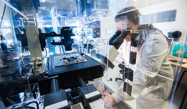
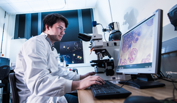
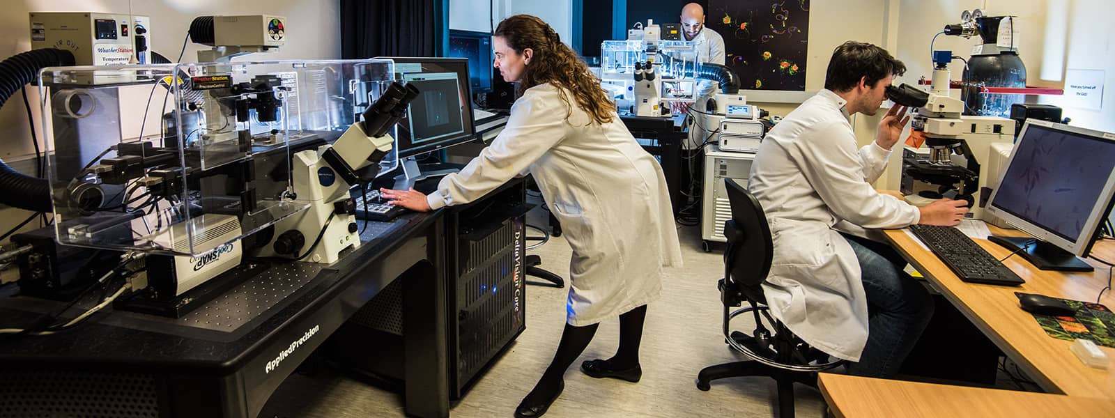
The BLS Bioimaging Facility houses various Fluorescent Light Microscopes including a DeltaVision deconvolution imaging system, a Crest X-light v3 Spinning Disc Confocal, a Leica Stellaris 5 and a Zeiss LSM880 with Airyscan module for near super-resolution imaging, all equipped with temperature and CO2 controlled chambers for live cell imaging and long time lapse experiments. The facility is also equipped with a fully-automated Zeiss AxioZoom.V16 stereozoom microscope with 15-color LED illumination and high-resolution optics with long working distances and extremely large fields of view.
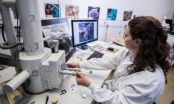
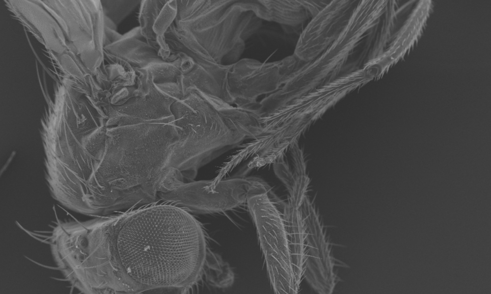
The facility has two Jeol electron microscopes: A Jeol JSM-5600 high vacuum Scanning Electron Microscope with tungsten filament, secondary election detector and digital output; and a Jeol JEM-1010, high-contrast Transmission Electron Microscope, with tungsten filament electron source and AMT CCD camera for digital image acquisition (bottom mounted).
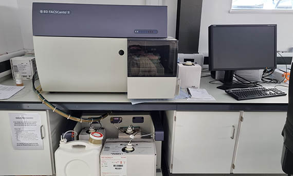
Flow cytometry facilities available include a Sony MA900 cell sorter with 4 lasers for detection of up to 14 parameters, and BD FACS Canto II or Beckman Coulter CytoFlex analysers, the later having facilities for 13 colour data acquisition and plate mode loading of samples from a 96 well plate.
Finally, the facility hosts a C-Trap Dymo300 automated optical-trap fluorescence microscope that enables direct correlation of molecular spatial dynamics with their physical interactions, funded by BBSRC ALERT 2024.